A Gram stain is a way of detecting bacteria and is often performed on the same sample as a sputum culture test. Gram-positive bacteria and gram-negative bacteriaThe name comes from the Danish bacteriologist Hans Christian Gram who developed the technique in 1884.

Pdf Mlt 415 Lab Report Gram Stain Techniques Muhamad Faizzudin Mohamad Zan Academia Edu
Start with an abstract.
. For agarose gels we recommend using Original GelRed Nucleic Acid Gel Stain or GelGreen Nucleic Acid Gel Stain. Sputum culture test vs. In a Gram stain different colored materials are applied to the sample which is then analyzed under a microscope.
This will allow the reader to see in short form the purpose results and. The Alternative 4 tube was used to inoculate a nutrient agar plate using the quadrant streak technique. That test tube only had a gram negative bacterium in it.
It is then possible to draw inferences. You will find more specific procedures for each biochemical test on the following pages. It is not enough to just identify your organism.
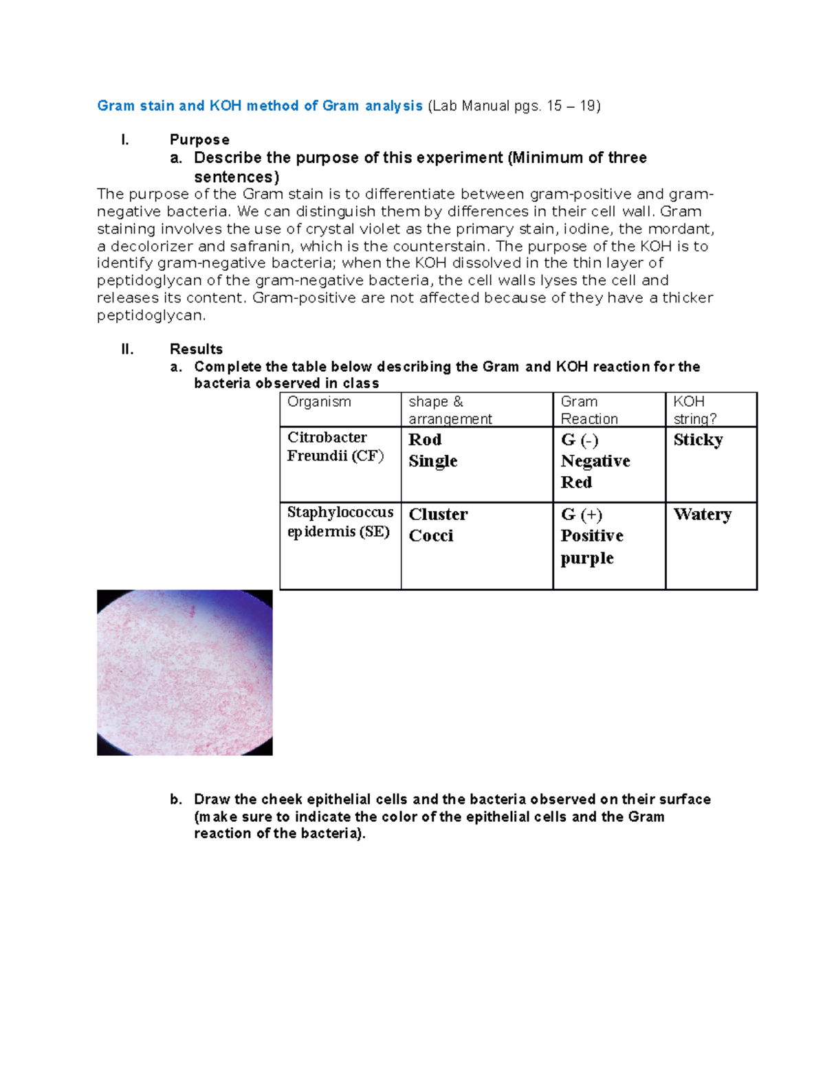
When doing a gram stain the results showed that the bacterium was Gram negative rods. It represents one aspect of water qualityIt is a microbiological analytical procedure which uses samples of water and from these samples determines the concentration of bacteria. For these reasons you will not find reference ranges for the majority of tests described on this web site.
This complex is a larger molecule than the original crystal violet stain and iodine and is insoluble in water. 1 A Gram stain from a carefully collected specimen with neutrophils and lancet-shaped diplococci staining gram-positive which are intracellular or encapsulated can provide strong support to the clinical diagnosis of pneumococcal pneumonia. Please see Table 2 for a complete list of each test purpose reagents.
Please consult your doctor or the laboratory that performed the tests to obtain the reference range if you do not have the lab report. The abstract is a very short summary of the paper usually no more than 200 words. GelRed in water is a newer safer formulation and our.
Part 1 of 3. Base the structure of your abstract on the structure of your paper. A Mannitol Salt Agar plate was obtained in order to test for mannitol fermentation.
GelRed 3X in water is ready-to-use for post-electrophoresis gel staining and is supplied in a 4L Cubitainer. You will need to look up the. Based on the change in color and the shape and size of cells the test can.
Conversely the the outer membrane of Gram negative bacteria is degraded and the thinner peptidoglycan layer of Gram negative cells is unable to retain the crystal violet-iodine complex and the color is lost. The gram negative bacterium grew on the agar and a gram stain was carried out. When you breathe you bring oxygen O 2 into your lungs and release carbon dioxide CO 2Carbon dioxide in your blood is present in three forms.
An SOP should be written for all procedures in the laboratory including specimen collection transport storage waste disposal Gram stain microscopy biochemistry measurements culture identification antimicrobial. Bacteriological water analysis is a method of analysing water to estimate the numbers of bacteria present and if needed to find out what sort of bacteria they are. The gram stain showed a gram negative rod was present.
We have included the basic procedure for doing each biochemical test in the table below. Results obtained by culture without evaluation for contamination may be noncontributory or misleading. Writing an Abstract and Introduction 1.
An inoculating loop was sterilized a sample of Unknown B was collected and a streak was made on the agar plate. The procedures are paraphrased from the National Committee for Clinical Laboratory Standards NCCLS 2000. You also need to know what antimicrobial agents your organism is susceptible to.
The lab report containing your test results should include the relevant reference range for your tests. More complete information on selective differential media can be obtained by consulting the Difco manuals in lab. This article explains the basic format of a lab report.
Higher concentrations of Original GelRed are available as 10000X in water or DMSO. Gram stain or Gram staining also called Grams method is a method of staining used to classify bacterial species into two large groups. The information presented in this lab is from The Manual of Clinical Microbiology 8th Ed.
SYTO 9 green fluorescent nucleic acid stain has been shown to stain live and dead Gram-positive and Gram-negative bacteria and it is a component of the LIVEDEAD BacLight Bacterial Viability Kits L-7007 L-7012 L-13152. Carbonic acid H 2 CO 3 CO 2 dissolved in blood and bicarbonate HCO 3- the predominant formBicarbonate is a negatively charged ion that is excreted and reabsorbed by your kidneys. Request forms report forms and other laboratory forms are all important components of the quality manual which documents the quality management system.
Gram staining differentiates bacteria by the chemical and physical properties of their. Next the MacConkey and EMB tests were performed.
Gram Stain And Koh Method Of Gram Analysis Report Gram Stain And Koh Method Of Gram Analysis Lab Studocu
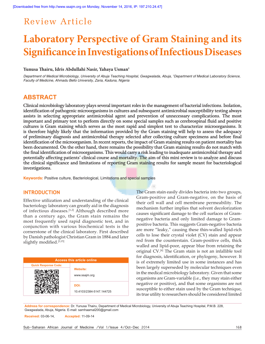
Pdf Laboratory Perspective Of Gram Staining And Its Significance In Investigations Of Infectious Diseases

Pdf Lab Report Of Microbiology 3 Akbar Haqi Academia Edu
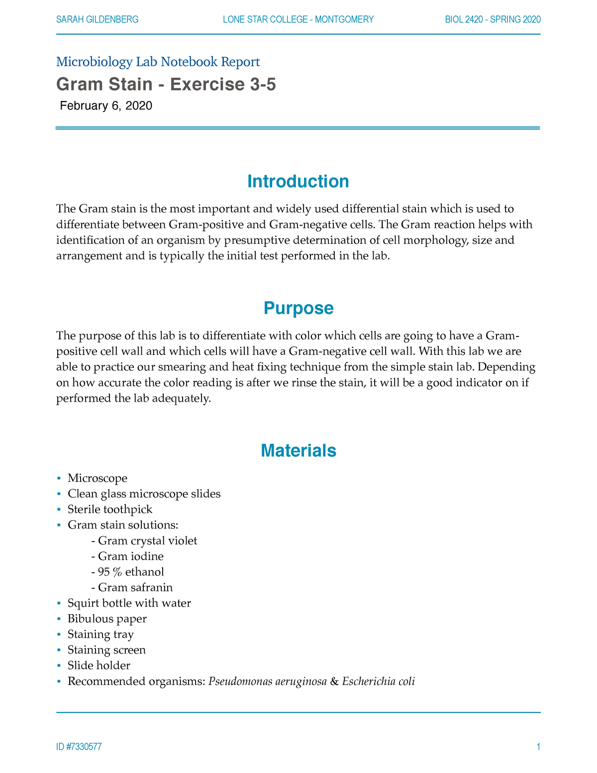
Complete Gram Stain Lab Microbiology Lab Notebook Report Gram Stain Exercise 3 February 6 2020 Studocu


0 Comments